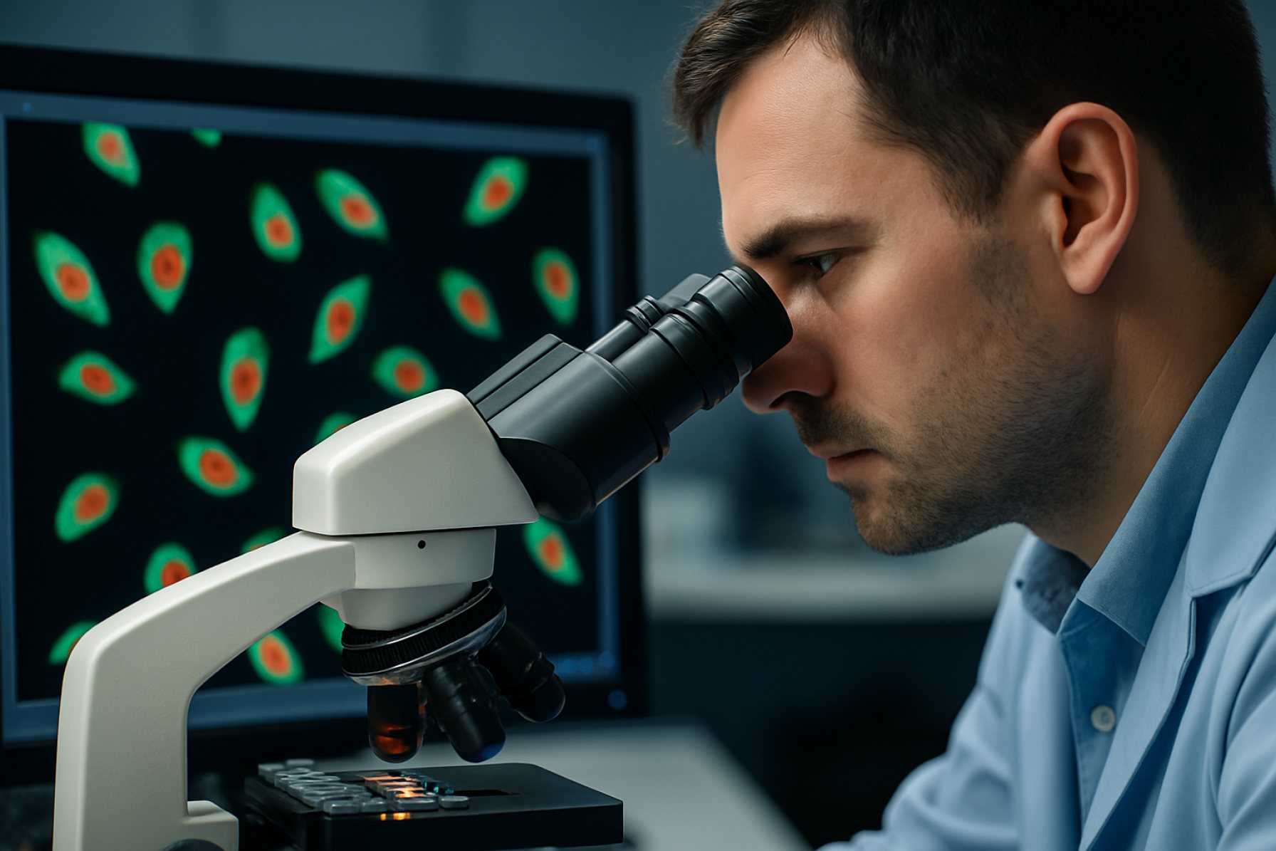
Single-Cell Imaging Technologies: The Game-Changer Transforming Our Understanding of Cellular Mysteries. Discover How Cutting-Edge Imaging Is Redefining Precision Medicine and Biological Research.
- Introduction: The Rise of Single-Cell Imaging
- Core Principles and Techniques in Single-Cell Imaging
- Breakthrough Innovations: From Fluorescence to Super-Resolution
- Applications in Disease Research and Precision Medicine
- Challenges and Limitations in Current Technologies
- Integration with Multi-Omics and Data Analysis
- Future Directions: AI, Automation, and Next-Gen Platforms
- Conclusion: The Impact and Promise of Single-Cell Imaging
- Sources & References
Introduction: The Rise of Single-Cell Imaging
Single-cell imaging technologies have revolutionized the study of cellular heterogeneity, enabling researchers to visualize and analyze the behavior, structure, and molecular composition of individual cells within complex tissues. Unlike traditional bulk analysis, which averages signals across populations, single-cell imaging provides spatial and temporal resolution at the level of individual cells, uncovering rare cell types, dynamic processes, and intricate cell-to-cell interactions that were previously obscured. This paradigm shift has been driven by advances in high-resolution microscopy, fluorescent labeling, and computational image analysis, allowing for the simultaneous measurement of multiple cellular features in situ.
The rise of single-cell imaging is closely linked to the growing recognition that cellular diversity underpins many biological phenomena, from embryonic development to disease progression. For example, in cancer research, single-cell imaging has revealed the presence of distinct subpopulations within tumors that may drive therapy resistance or metastasis. In neuroscience, these technologies have enabled the mapping of neural circuits with unprecedented detail, shedding light on the cellular basis of behavior and cognition. Furthermore, the integration of imaging with other single-cell approaches, such as transcriptomics and proteomics, is providing a more comprehensive understanding of cell states and functions.
As single-cell imaging technologies continue to evolve, they are poised to play a central role in precision medicine, drug discovery, and systems biology. Ongoing innovations in imaging hardware, molecular probes, and data analysis are expanding the scale, speed, and depth of single-cell investigations, promising new insights into the complexity of life at the cellular level (Nature Methods; Cell).
Core Principles and Techniques in Single-Cell Imaging
Single-cell imaging technologies are grounded in the ability to visualize and quantify molecular and structural features at the resolution of individual cells, enabling the dissection of cellular heterogeneity within complex tissues. The core principles involve high spatial and temporal resolution, sensitivity to low-abundance targets, and minimal perturbation to native cellular states. Central to these technologies are advanced microscopy techniques, such as confocal and two-photon microscopy, which provide optical sectioning and deep tissue penetration, respectively. Super-resolution methods, including STED, PALM, and STORM, surpass the diffraction limit of light, allowing visualization of subcellular structures at nanometer scales (Nature Methods).
Fluorescent labeling is a cornerstone technique, utilizing genetically encoded fluorescent proteins or synthetic dyes to tag specific proteins, nucleic acids, or organelles. Multiplexed imaging approaches, such as spectral unmixing and sequential hybridization, enable simultaneous detection of multiple targets within the same cell (Cell Press). Live-cell imaging techniques, often combined with microfluidics, allow dynamic monitoring of cellular processes in real time, providing insights into cell signaling, division, and migration.
Quantitative image analysis, powered by machine learning and artificial intelligence, is increasingly essential for extracting meaningful data from high-dimensional single-cell images. These computational tools facilitate cell segmentation, feature extraction, and phenotypic classification, driving discoveries in developmental biology, cancer research, and immunology (Nature Methods). Collectively, these core principles and techniques underpin the transformative potential of single-cell imaging in biomedical research.
Breakthrough Innovations: From Fluorescence to Super-Resolution
The evolution of single-cell imaging technologies has been marked by a series of transformative innovations, particularly the transition from conventional fluorescence microscopy to advanced super-resolution techniques. Traditional fluorescence microscopy, while invaluable for visualizing cellular structures and protein localization, is fundamentally limited by the diffraction barrier, restricting resolution to approximately 200 nanometers. This limitation has historically impeded the detailed study of subcellular processes and molecular interactions within individual cells.
The advent of super-resolution microscopy—encompassing methods such as Stimulated Emission Depletion (STED), Photoactivated Localization Microscopy (PALM), and Stochastic Optical Reconstruction Microscopy (STORM)—has shattered this barrier, enabling visualization at resolutions down to 20 nanometers or less. These breakthroughs have allowed researchers to observe the spatial organization of proteins, nucleic acids, and organelles with unprecedented clarity, revealing previously inaccessible details of cellular architecture and dynamics. For instance, super-resolution imaging has elucidated the nanoscale arrangement of synaptic proteins in neurons and the organization of chromatin domains in the nucleus, providing critical insights into cellular function and disease mechanisms (Nature Methods).
Moreover, the integration of super-resolution techniques with live-cell imaging and multiplexed labeling strategies has further expanded the capabilities of single-cell analysis. These advances facilitate real-time tracking of molecular events and the simultaneous visualization of multiple targets, offering a comprehensive view of cellular heterogeneity and dynamic processes (Cell). As a result, the leap from fluorescence to super-resolution represents a pivotal milestone, driving forward our understanding of cell biology at the single-cell level.
Applications in Disease Research and Precision Medicine
Single-cell imaging technologies have revolutionized disease research and precision medicine by enabling the visualization and quantification of molecular and cellular heterogeneity at unprecedented resolution. In oncology, these technologies allow researchers to dissect tumor microenvironments, track clonal evolution, and identify rare cell populations responsible for drug resistance or metastasis. For example, multiplexed imaging platforms such as cyclic immunofluorescence and imaging mass cytometry can simultaneously map dozens of protein markers within individual tumor cells, providing insights into spatial organization and cell-to-cell interactions that drive disease progression Nature Reviews Genetics.
In immunology, single-cell imaging has been instrumental in characterizing immune cell diversity and function within tissues, revealing how specific cell subsets contribute to autoimmune disorders or respond to infections. These insights have informed the development of targeted immunotherapies and vaccines tailored to individual patient profiles Cell.
Furthermore, in the context of precision medicine, single-cell imaging technologies facilitate the identification of biomarkers predictive of therapeutic response or disease outcome. By integrating imaging data with genomic and transcriptomic analyses, clinicians can stratify patients more accurately and design personalized treatment regimens. The ability to monitor dynamic cellular responses to drugs in real time also supports adaptive treatment strategies, minimizing adverse effects and improving efficacy Nature Medicine.
Overall, single-cell imaging technologies are driving a paradigm shift in disease research and clinical practice, enabling a deeper understanding of pathophysiology and supporting the realization of truly individualized medicine.
Challenges and Limitations in Current Technologies
Despite remarkable advances, single-cell imaging technologies face several significant challenges and limitations that impact their widespread application and data interpretation. One major hurdle is the trade-off between spatial resolution, temporal resolution, and imaging depth. High-resolution techniques, such as super-resolution microscopy, often require longer acquisition times and are limited in their ability to penetrate deep into tissues, restricting their use in live or thick biological samples (Nature Methods). Additionally, phototoxicity and photobleaching remain persistent issues, especially during prolonged imaging sessions, potentially altering cellular physiology and compromising data integrity.
Another limitation is the complexity and cost of advanced imaging platforms. Many state-of-the-art systems require specialized equipment and expertise, making them less accessible to standard laboratories (Cell). Furthermore, the vast amount of data generated by single-cell imaging necessitates robust computational tools for storage, processing, and analysis. Current algorithms may struggle with the high dimensionality and heterogeneity of single-cell data, leading to challenges in accurate segmentation, tracking, and quantification (Nature Biotechnology).
Finally, multiplexing—the ability to simultaneously visualize multiple molecular targets—remains limited by spectral overlap and the availability of suitable probes. This constrains the depth of biological insight that can be achieved in a single experiment. Overcoming these challenges will require continued innovation in imaging hardware, probe chemistry, and computational analysis to fully realize the potential of single-cell imaging technologies.
Integration with Multi-Omics and Data Analysis
The integration of single-cell imaging technologies with multi-omics approaches has revolutionized our ability to dissect cellular heterogeneity and function at unprecedented resolution. By combining high-content imaging with genomics, transcriptomics, proteomics, and metabolomics, researchers can correlate spatial and morphological features with molecular profiles in individual cells. This synergy enables the identification of rare cell types, dynamic cellular states, and intricate cell-cell interactions within complex tissues. For instance, spatial transcriptomics platforms now allow the mapping of gene expression patterns directly onto tissue sections, while advanced imaging mass cytometry can quantify dozens of proteins simultaneously at subcellular resolution (Nature Methods).
However, the integration of these diverse data types presents significant analytical challenges. Data from imaging and omics platforms differ in scale, dimensionality, and noise characteristics, necessitating sophisticated computational frameworks for alignment, normalization, and interpretation. Machine learning and artificial intelligence are increasingly employed to extract meaningful patterns, perform cell-type classification, and reconstruct spatially resolved molecular networks (Cell). Open-source tools and standardized pipelines are being developed to facilitate reproducible analysis and data sharing across laboratories (Human Cell Atlas).
As these integrative strategies mature, they promise to yield comprehensive atlases of tissue organization and disease progression, ultimately informing precision medicine and therapeutic development. The continued evolution of single-cell imaging and multi-omics integration will be pivotal for unraveling the complexity of biological systems at the single-cell level.
Future Directions: AI, Automation, and Next-Gen Platforms
The future of single-cell imaging technologies is being shaped by the integration of artificial intelligence (AI), automation, and next-generation platforms, promising to revolutionize both the scale and depth of cellular analysis. AI-driven image analysis algorithms are increasingly capable of extracting complex, high-dimensional features from vast imaging datasets, enabling the identification of subtle phenotypic variations and rare cell states that would be challenging to discern manually. For instance, deep learning models can now automate cell segmentation, classification, and tracking with unprecedented accuracy, reducing human bias and accelerating data interpretation (Nature Methods).
Automation is further enhancing throughput and reproducibility in single-cell imaging. Robotic sample handling, automated microscopy, and integrated data pipelines are streamlining workflows, making it feasible to image and analyze thousands to millions of cells in a single experiment. This scalability is crucial for large-scale studies, such as drug screening or tissue atlasing, where statistical power and consistency are paramount (Cell).
Next-generation platforms are also emerging, combining advanced optics, microfluidics, and multiplexed labeling strategies. These systems enable simultaneous imaging of multiple molecular targets and dynamic cellular processes at high spatial and temporal resolution. The convergence of these innovations is expected to unlock new biological insights, such as mapping cellular heterogeneity in complex tissues and understanding dynamic cell-cell interactions in real time (Nature Biotechnology). As these technologies mature, their integration with cloud-based analytics and open data standards will further democratize access and accelerate discovery in single-cell biology.
Conclusion: The Impact and Promise of Single-Cell Imaging
Single-cell imaging technologies have fundamentally transformed our understanding of cellular heterogeneity, enabling unprecedented insights into the spatial and temporal dynamics of individual cells within complex tissues. By allowing researchers to visualize and quantify molecular events at the single-cell level, these technologies have revealed the intricate variability that underlies development, disease progression, and therapeutic response. The impact of single-cell imaging is particularly evident in fields such as cancer biology, immunology, and neuroscience, where cellular diversity plays a critical role in function and pathology. For instance, the ability to track cell fate decisions and signaling pathways in real time has led to the identification of rare cell populations and novel biomarkers, informing both basic research and clinical applications Nature Reviews Genetics.
Looking forward, the promise of single-cell imaging lies in its continued integration with other high-throughput single-cell technologies, such as transcriptomics and proteomics, to provide a more comprehensive, multi-dimensional view of cellular states. Advances in imaging resolution, multiplexing capacity, and computational analysis are expected to further enhance the sensitivity and scalability of these approaches, making it possible to map entire tissues and organs at single-cell resolution Cell. As these technologies become more accessible and standardized, their adoption in both research and clinical settings will likely accelerate, driving new discoveries and enabling more precise diagnostics and personalized therapies. Ultimately, single-cell imaging stands as a cornerstone of modern cell biology, poised to unlock deeper understanding of life at its most fundamental level.
